
Keywords
parasitic disease
nasal
ulcerative-budding lesions
sandfly
How to Cite
Abstract
Introduction :
Cutaneous leishmaniasis is a parasitic skin disease caused by a parasite of the Leishmania group, transmitted to humans by the bite of a blood-sucking insect, the sandfly (phlebotomus). Some of its clinical forms can be mutilating. The objective of this work was to report a case of cutaneous leishmaniasis with nasal localization in its ulcerocrustous and budding form, diagnosed in a patient and managed at the Dermatology-Venereology department of the Niamey National Hospital.
Observation :
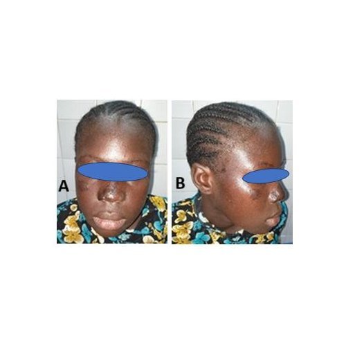
We report the case of a 16-year-old female patient, residing in a rural area, presenting with ulcerocrustous and budding, painless, edematous lesions with nasal deformation. A smear of the dermal fluid performed on the patient revealed leishmania. The diagnosis of cutaneous leishmaniasis was thus confirmed. This form with nasal localization can be mistaken for verrucous cutaneous tuberculosis, rhinophyma, or a basal cell carcinoma. Management with metronidazole allowed for complete healing of the lesions, leaving, however, an indelible and unaesthetic hyperpigmentation.
Conclusion :
Cutaneous leishmaniasis is a common tropical parasitic disease in Niger. The ulcerocrustous forms can sometimes be mutilating. Adequate management with antibiotics yields satisfactory results.
References
1. Salissou L, Brah S, Sani Maman L, Daou M, Assane F, Balaka A, et al. Traitement de la leishmaniose cutanée par le métronidazole : à propos de 78 cas au Niger. J Rech Sci Univ Lomé. 2016;18(1):151–6. doi:10.4314/jrsul.v18i1.
2. Mokni M. Leishmanioses cutanées (Cutaneous leishmaniasis). Ann dermatol venereol. 2019; 146 (3): 232 – 246.
3. Develoux M, Blanc L, Garba S, Mamoudou H, Ravisse P, Cenac A. Étude clinique et épidémiologique de la leishmaniose cutanée au Niger. Cah Etudes Rech Francoph Santé. 1991;1(2):130–4..
4. Soares VB, Almeida AS, Sabroza PC, Vargas WP. Epidemiological surveillance of tegumentary leishmaniasis: local territorial analysis. Rev Saude Publica. 2017 ; 26;51:51. doi: 10.1590/S1518-8787.2017051006614.
5. Blaizot R, Lamine MM, Saout M, Issa K, Laminou IM, Duvignaud A, Demar M, Doutchi M. Molecular detection of Leishmania major human infections in the Zinder area, Niger. BMC Infect Dis. 2025 Jul 1;25(1):871. doi: 10.1186/s12879-025-11229-2.
6. Gilkey T, Ulman CA, McGwire B, Plaza JA. Cutaneous Leishmania aethiopica diagnosed in the United States. Dermatol Online J. 2021 Aug 15;27(8). doi: 10.5070/D327854705.
7. Qasmi S, Elguelbazouri N, Belgnaoui FZ, Marcil T, Bouhllab J, Senouci K, et al. Childhood cutaneous leishmaniasis: Experience of a Moroccan unit of dermatology. Dermatol Online J. 2008 Dec 15;14(12):18.
8. Gilkey T, Ulman CA, McGwire B, Plaza JA. Cutaneous Leishmania aethiopica diagnosed in the United States. Dermatol Online J. 2021 Aug 15;27(8). doi: 10.5070/D327854705.
9. El Kartouti A, Elbenaye J, Miloudi M. Leishmaniose cutanée érysipéloide: à propos d’une observation clinique. Pan Afr Med J. 2015 May 25;21:54. doi:10.11604/pamj.2015.21.54.5967.
10. Doulla M., Salissou L., Korsaga/Some N., Ouédraogo M., Aminou L., Zaki H., et al. (2022). Warty cutaneous tuberculosis of the nose: A rare localization. Our Dermatol Online. 2022;13(1):50– 52. doi:10.7241/ourd.20221.11..
11. Bari AU, Ejaz A. Rhinophymous leishmaniasis: A new variant. Dermatol Online J. 2009 Feb;15(3):10. doi:10.5070/D36GG453Q7.
12. Chauhan R, Loewenstein SN, Hassanein AH. Rhinophyma: Prevalence, Severity, Impact and Management. Clin Cosmet Investig Dermatol. 2020; 11;13:537-551. doi: 10.2147/CCID.S201290.
13. Ramli I, Hassam B. Cas historique de carcinome basocellulaire du nez. Pan Afr Med J. 2014, 27;19:326. doi:10.11604/pamj.2014.19.326.4393.
14. Salissou L, Doulla M, Ouedraogo MM, Brah S, Daou M, Ali D, et al. Lésion ulcérocroûteuse de l’angle interne de l’œil droit: la leishmaniose cutanée en est une cause. Our Dermatol Online. 2018;9(2):203–206. doi:10.7241/ourd.20182.26.
Metrics
Downloads

This work is licensed under a Creative Commons Attribution 4.0 International License.
Copyright (c) 2025 S Laouali, M Laouali Idi, M Ouédraogo Mamadou, S Ousmane, R Halidou Sidibé, A Inouss Issa, A Abdourahamane, A Ango, M Abdoulaye, M Harouna