

Keywords
histological type
prognosis
Niamey
Hospital
How to Cite
Abstract
Introduction :
Uterine cancers are a heterogeneous group of malignant tumors located in the cervix and/or the body of the uterus. Histologically, they include epithelial tumors, mesenchymal tumors, and mixed tumors. The aim of this study was to describe the histological profile of uterine cancers diagnosed at the Laboratory of Anatomy and Pathological Cytology of the National Hospital of Niamey.
Methodology :
This is a three-year cross-sectional study with retrospective data collection that included all histologically diagnosed uterine cancers. The variables studied included patient age and sex, lesion location, histological type, tumor stage, and patient outcome. Pearson's Chi-square test and Student-Fisher test were used to investigate relationships between variables, with a significance threshold set at P < 0.05.
Results :
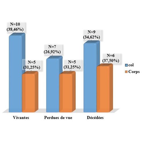
During the study period, 42 cases of uterine cancer were collected out of a total of 89 gynecological cancers, representing an overall frequency of 47.20%. In 61.90% of cases (N=26), the cancer was located in the cervix. The average age was 55.19 years (range : 32–81 years), varying from 53.32 years for cervical cancer to 58.12 years for uterine body cancer. The most common histological group was carcinoma, accounting for 75.53% of cervical cases and 26.47% of body cases. Furthermore, out of 8 sarcoma cases, 7 (87.50%) were found in the uterine body. Squamous cell carcinoma was the most common histological type in the cervix (76.92%), while carcinosarcoma was the most prevalent in the body (25%). Tumors classified as pT1 were the most frequent in both the cervix (69.24%) and the body (62.5%). The mortality rates for cervical and uterine body cancers were 34.62% and 37.50%, respectively.
Conclusion :
Uterine cancer predominantly affects postmenopausal women and is mainly located in the cervix, with carcinoma being the dominant histological group. The mortality rate is similar for both cervical and uterine body cancers, affecting more than a quarter of the patients.
References
1. Sancho-Garnier H. Epidémiologie des cancers gynécologiques : utérus, ovaire, vulve et vagin. EM consulte. 2013 ; 2(4): 85-99.
2. Stewarty BW, Kleihues P. Les cancers de l’appareil génital féminin. In : Le cancer dans le monde. IARC Press, Editor. Lyon. 2005 : 218-26.
3. World Health Organization (WHO). Classification of Tumours Female Genital Tumours: 5th Edition. 2020: 270p.
4. Nayama M, Nouhou H, Souna M K, Idi N, Garba M, Tahirou A et al. Cancers gynecologiques et mammaires: aspects epidemiologiques et histologiques dans le service d’anatomie et cytologie pathologique de la faculte des sciences et de la sante de niamey niger. Mali Médical. 2006 T XXI N° 3 : 43-49.
5. Amégbor K, Alfa A K, Darré T, Napo K G A, Akpadza K. Aspects épidémiologiques et histopathologiques des cancers gynécomammaires au Togo. Med Trop. 2011 ; 71 : 451-453.
6. Echafi Y. Cancer du col utérin : Etude anatomopathologique. Thèse de médecine. Université Cadi Ayyad. 2019: 148p.
7. Bannour N, Boughizane S, Naifer R. Le cancer invasif du col utérin dans le centre tunisien. Approches épidémiologiques, clinique et thérapeutique. Étude rétrospective de 96 cas. 2004 ; 6 (7) ; 481 – 488.
8. Ouédraogo A S, Sanou L A, Ramde W N, Konsegre V, Franck I , Lompo G M et al. Aspects histoépidémiologiques du cancer de l’endomètre. J. Afr. Cancer. 20113(4), 251255. Doi:10.1007/s12558-011-0191-8
9. Majda BI. Tumeurs de l’endomètre type II. Thèse Médecine. Université Cadi Ayyad de Marrakech. 2018. N°038. 36p.
10. Sando Z, Tsuala F J, Ymele F F, Hortence F J, Telesphore M E, Oyono E JL . Profil des cancers gynécologiques et mammaires à Yaoundé Cameroun. Pan Afr Med J. 2014;17(28): 34-47.
11. Berraho M, Bendahhou K, Obtel M, Zidouh A, Benider A, Errihani H et al. Cervical cancer in morocco: Epidemiological profile from two main oncological centers. Asian Pac J Cancer Prev. 2012;13(7): 3153-7.
12. Sawadogo TA. Diagnostic anatomopathologique du cancer du col de l’uterus au centre hospitalier universitaire souro sanou de bobo dioulasso. License en biologie. Université polytechnique de BoboDioulasso. 2016 ; 30p.
13. Raja A. Prise en charge laparoscopique des cancers de l’endomètre. Thèse médecine. Université Cadi Ayyad de Marrakech. 2014. N°50.
14. Chang YN, Zhang Y, Wang YJ. Effect of hysteroscopy on the peritoneal dissemination of endometrial cancer cells: a metaanalysis. Fertil Steril. 2011; 96(4): 957– 961.
15. Noer MC, Antonsen SL, Ottesen B, Christensen IJ, Høgdall C. Type I Versus Type II Endometrial Cancer: Differential Impact of Comorbidity. Int J Gynecol Cancer. 2018 Mar;28(3):586-593. doi: 10.1097/IGC.0000000000001184
16. Brinton LA, Felix AS, McMeekin DS, Creasman WT, Sherman ME, Mutch D, et al . Etiologic heterogeneity in endometrial cancer: evidence from a Gynecologic Oncology Group trial. Gynecol Oncol. 2013 May;129(2):277-84. doi: 10.1016/j.ygyno.2013.02.023.
17. Zannoni GF, Vellone VG, Arena V, Prisco MG, Scambia G, Carbone A, et al. Does high-grade endometrioid carcinoma (grade 3 FIGO) belong to type I or type II endometrial cancer? A clinical-pathological a n d i m m u n o h i s t o - c h e m i c a l study. Virchows ArchInt J Pathol. 2010; 457:27–34.
18. Wang Y, Sun Y, Sun F, Han P, Fan R, Ren F. Comparison of clinical characteristics and prognosis between type I and type II endometrial cancer: a single-center retrospective study. Discov Oncol. 2023 Nov 23;14(1):211. doi: 10.1007/s12672-023-00820-1
19. Schlosshauer PW, Pirog EC, Levine RL, Ellenson LH. Mutational analysis of the CTNNB1 and APC genes in uterine endometrioid carcinoma. Mod Pathol. 2000; 13:1066–71.
20. Diarra IM. Aspects clinique et thérapeutique de cancer du col de l’utérus dans le service de gynécologie et d’obstétrique du CHU du point G. Thèse de médecine. Université des Sciences, des Techniques et des Technologies de Bamako 2009, N° 150 : p48-52.
21. Samaké S. Aspects épidémiologiques, anatomo–pathologiques, cliniques et thérapeutiques des cancers du col utérin. Thèse médecine Bamako, 2014 : N°111, p75.
Metrics
Downloads

This work is licensed under a Creative Commons Attribution 4.0 International License.
Copyright (c) 2025 I Boubacar, B Efared, I Seriba Coulibaly, A Koussou Moussa, H Salifou Boureima, A Balaraba Abani, G Mahamadou, M Nayama, H Nouhou